Patient Information:
A nineteen year old female patient was referred with bilateral metatarsalgia for a joint podiatry and orthopaedic opinion.
Symptoms:
The young patient presented with chronic plantar forefoot pain, lasting over 4 years described as "like walking on hot stones." The pain was aggravated by standing and walking; it was limiting her activities of daily life. The patient had no medical conditions other than obesity, a body weight of 136 kg (BMI 58.8). Her pain severity was measured on a Visual Analog Scale for Pain (VAS) to be 8/10 standing and 10/10 when trying to walk, causing a limp (antalgic gait).
Diagnosis and Imaging Investigations:
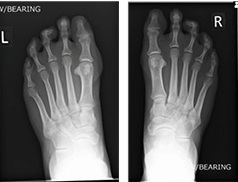 Weight-bearing X-ray (Left) and (Right)
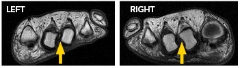 T1 weighted short axis foot MRI
The symptoms were attributed to overload metatarsalgia due to congenitally short first metatarsal bones in both feet. MRI and X-ray investigations can be seen at the right. The report verified there was no bone or joint signal typical of bone stress (or synovitis). Signal consistent with inter-metatarsal bursitis between the 2nd to 3rd inter-metatarsal space (indicated by yellow arrows). X-rays show very slight clawed second toes.
Pressure Measurement Evaluation:
A plantar pressure assessment using the F-Scan™ System was performed for a better understanding of the foot function and pressures. The purpose of the gait assessment using F-Scan System was to test and optimize the benefits of the orthotic device; and if this failed, determine if forefoot surgery was necessary and what affect this may have on forefoot pressures.
Outcome:
As a result of the plantar pressure measurements, the orthotic devices utilized were modified to optimize pressure redistribution at the forefoot. This was achieved by adding a forefoot rocker to the orthoses. Previous devices included a casted ¾ Functional Foot Orthotic (FFO) and variations of a pre-fabricated orthoses with metatarsal bar were not as successful. Patient showed an immediate increase in comfort and improved gait symmetry; as well as at the initial follow-up visit. At pre-surgical follow-up after 3 months, pain scores reduced to 4/10, so the patient opted not to have the surgery; therefore saving the trust a substantial cost. Follow-up plantar pressure scans were not needed due to good clinical response. In addition, the patient has taken the public health message on-board to reduce her foot pressure by loosing body weight, which we think the visual feedback of the pressure assessment aided in.
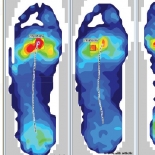
Walking Plantar Pressure Measurement:
Screenshot below shows peak pressure at the site of pain and pathology: Left 2nd metatarsal (met) head 815 KPa, Right 2nd met head 930 KPa, additionally there was little to no toe contact.
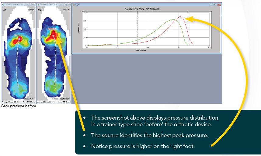
Peak Pressure Comparison
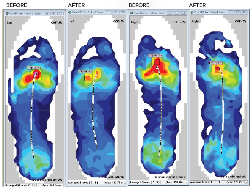
The screenshot to the right displays pressure distribution in a trainer type shoe only "before" and with the orthotic device "after."
Notice how the pressure has been re-distributed in the "after" image, where there is reduced heel pressure, increased midfoot pressure, reduced forefoot pressure and increased toe pressure.
Peak pressure, located at the forefoot, under the 2nd metatarsal head at the site of pain, is lower in the left foot by 24% and on the right foot by 30% when wearing the optimized foot orthoses.
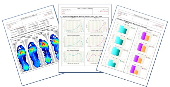
Comparative Reports As an Educational Tool:
Peak pressure reports generated with the click of a button, provide quick comparison, aid in patient education and document treatment progress.
 Peak pressure reports are generated at the click of a button and provide a way to document treatment progress.
About the Clinician:
Dr. Jill Halstead-Rastrick (PhD) is the Principal Podiatrist in Biomechanics, Orthotics and Footwear at Salford Royal Hospital (NHS) Foundation Trust. She is also a visiting research fellow at the University of Leeds and is the co-founder of MSKUK the special advisory group for Podiatrists.
Acknowledgments:
I would like to thank Mr. Ian Reilly for reviewing this report and providing helpful suggestions regarding the presentation of the XRrays and MRI.
Learn how Tekscan systems deliver actionable gait analysis data.
Read About our Gait Analysis Systems