 Dr. Sangiv I. Patel (DDS, RDH, AFAAID)Patient Overview
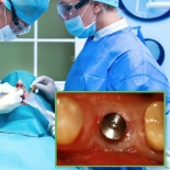
A 56-year-old male patient with a recent history of a lower right first molar that had fractured and was deemed unrestorable.
- The patient was advised by his periodontist to do a dental implant (not another bridge) in order to regain function.
- The molar was consequentially removed and the site received a bone graft.
- A dental implant was placed by the periodontist after the bone graft healed.
The patient knew about our facility and advanced dental technology, including CEREC CAD/CAM and T-Scan technology. Dr. Patel’s care, in conjunction with the application of these technologies, was the primary reason for the patient selecting his practice for his restorative care.
Based on the pre-restorative Center of Force (COF) measured by T-Scan, it is very possible this patient would have never lost that particular molar if the COF was managed with an equilibration prior to the molar fracturing.
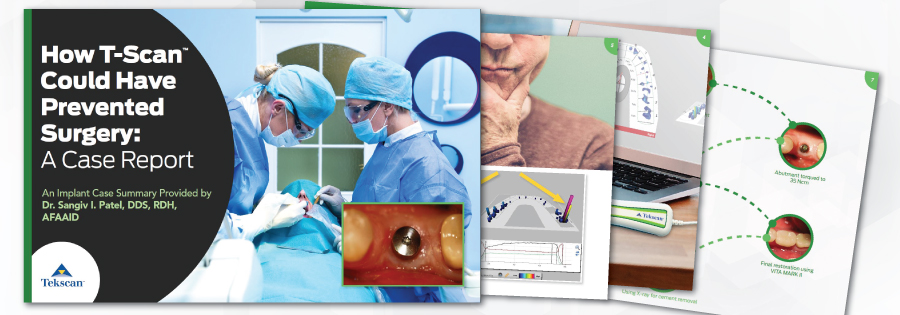
For More Ways to Use T-Scan in Implant Applications,