 In this case, we’ll follow a 61-year-old male patient who’s an executive with a high-stress job.
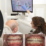
- His four front teeth were loose—they wiggle a lot.
- There was no sign of inflammation or decay, and while he does have a few fillings here and there, nothing stands out as an obvious tooth issue.
- There’s a faint black line around the root of the tooth called the periodontal ligament—it holds the tooth into the bone. When you compare it to the bottom opposing teeth, there is no widening of the periodontal space. These teeth are not mobile; they’re solid as a rock.
- The spaces around the front four teeth are filled with elastic tissue, so these teeth do move. In fact, when the patient bites his teeth together, those upper teeth push outward. This indicates there is fremitus.
Teeth #7-10 demonstrated Class 3 mobility which means they’re depressible in the socket. If you pushed his front teeth upwards, you could actually push them into his skull. That’s how mobile these four teeth are! Obviously, there is a problem...
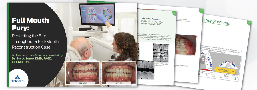
Click to Download (11MB)